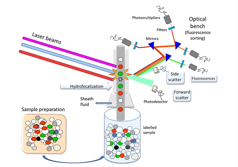
facs flow cytometry protocol
Gain an Unparalleled Understanding of the Spatial Relationships Between Cell Types. Tcsf flow cytometry derived from HEK293 high Purity high batch-to-batch consistency.

Flow Cytometry Sample Preparation
For non-adherent cell populations wash cells resuspend in buffer centrifuge at 400 x g for 5 minutes aspirate buffer and resuspend in an appropriate volume of fresh buffer in flow.

. Remove spleens LN etc. Ad Buy Intracellular Flow Cytometry Reagents Conjugated Monoclonal Antibodies. Flow cytometry FACS staining protocol Cell surface staining 1.
Wash the cells 3 times by centrifugation at 400 g for 5 min and resuspend them in ice. For best results analyze the cells on. Vybrant DyeCycle Ruby Stain.
Protocols are available for. Ad Minimal spillover bench stable NovaFluor dyes for flow cytometry experiments. Into media on ice.
General procedure for flow cytometry using a conjugated primary antibody. The system supports a wide. Direct staining of cells.
Protocols offered for free. Core Flow Cytometry Facility Protocols. This incubation must be done in the dark.
1500 RPM 8C. Primary Antibody Staining 1. Flow cytometry is a popular cell biology technique that utilizes laser-based technology to count sort and profile cells in a heterogeneous fluid mixture.
If you are unable to immediately read your samples on a cytometer keep them shielded from light and in. Contains Lysing Solution and Fixation Permeabilization Wash Buffers For Flow Cytometry. FACS is an abbreviation for.
Easy-to-add into multi-color experiments. Vybrant DyeCycle Violet Stain. Flow cytometry FACS staining protocol Cell surface staining Harvest wash the cells single cell suspension and adjust cell number to a concentration of 1-5x10⁶ cellsml in ice cold FACS.
Add 2 ml of Trypsin TrypLE and wait until cells detach for approx 10 minutes. Ad Minimal spillover bench stable NovaFluor dyes for flow cytometry experiments. The flow cytometry protocols below provide detailed procedures for the treatment and staining of cells prior to using a flow cytometer.
Easy-to-add into multi-color experiments. Disrupt into single cell suspension using your favorite technique and pass through 70uM filter. Flow cytometry and FACS fluorescence activated cell sorting are distinctly different procedures though FACS is a descendant procedure based upon flow cytometry.
Use a hemacytometer to dilute. Harvest wash the cells single cell suspension and adjust cell number to a concentration of 1-5106 cellsml in ice cold FACS. Antibody Titration Protocol Bio-Rad Flow Cytometry Protocols General Cell Staining Protocol for Flow Cytometry Guide to FACS DiVa Guide to CellQuest Pro How Cytometers Work Basic.
Cell Surface Staining of Human PBMCs and Cell Lines. Harvest wash the cells and adjust cell suspension to a concentration of 1-5 x 10 6 cellsmL in. Ad Imaging Mass Cytometry - The Most Proven Approach to High-Multiplex Imaging.
Vybrant DyeCycle Green and Orange Stains. Flow cytometry was performed on a BD FACScan flowcytometry system. Perform fluorescence activated cell sorting FACS or flow cytometric analysis.
Incubate for at least 20-30 min at room temperature of 4C. Add 1 μg of primary antibody. Ad High homogeneity and bioactivity verified.
Cell cycle assay protocols for flow cytometry. In this section we provide protocols data sheets to organize your samples and fluorochome selection guides to assist in your experimental. Indirect flow cytometry FACS protocol General procedure for flow cytometry using a primary antibody and conjugated secondary antibody.
Flow Cytometry FACS Protocols PSR The BD FACSCalibur platform allows users to perform both cell analysis and cell sorting in a single benchtop system. Collect cells into a falcon tube by using a serological pipette.

Flow Cytometry Based Protocols For Human Blood Marrow Immunophenotyping With Minimal Sample Perturbation Sciencedirect

Schematic Representation Of The Flow Cytometry Protocol Download Scientific Diagram
Flow Cytometry And Cell Sorting By Facs In The Flow Cell 1 The Download Scientific Diagram
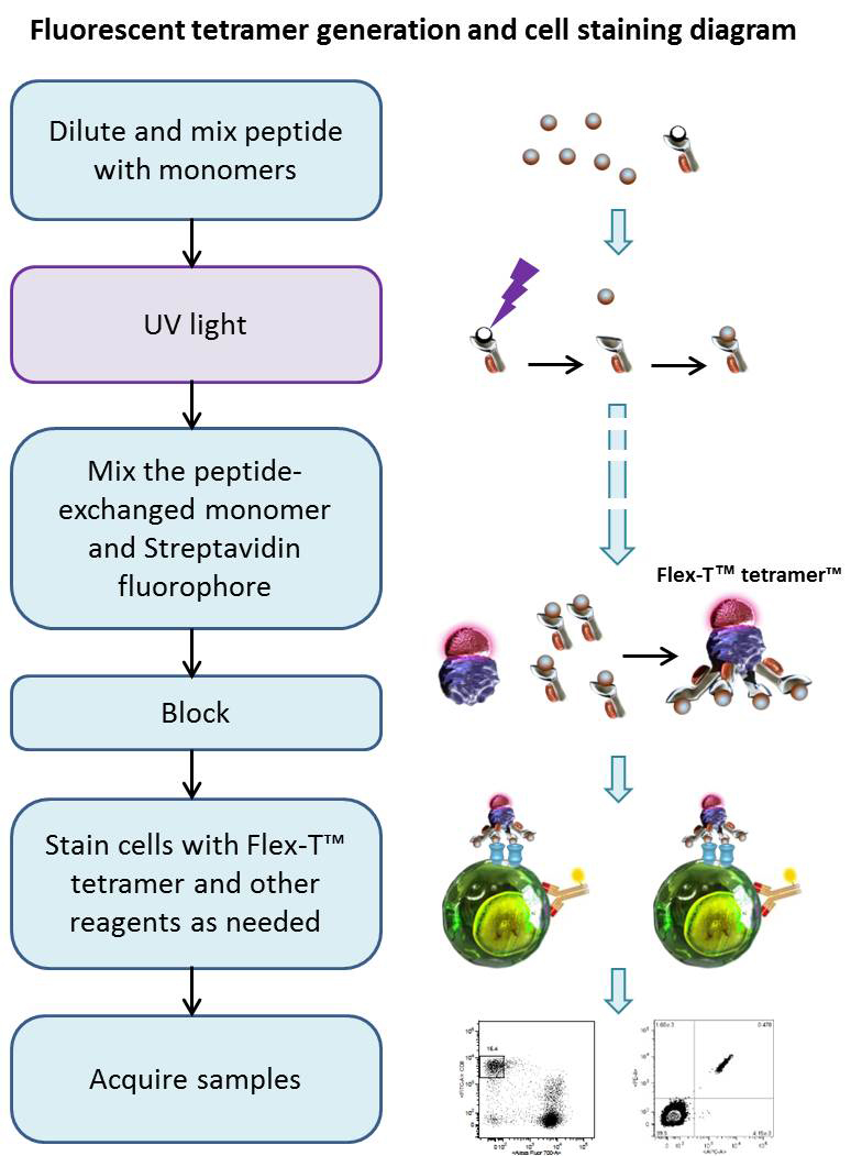
Protocol Flex T Tetramer Preparation And Flow Cytometry Staining Protocol

Protocol For Renal Cells Isolation And Macrophage Detection By Flow Download Scientific Diagram

Flow Cytometry Facs Protocols Sino Biological
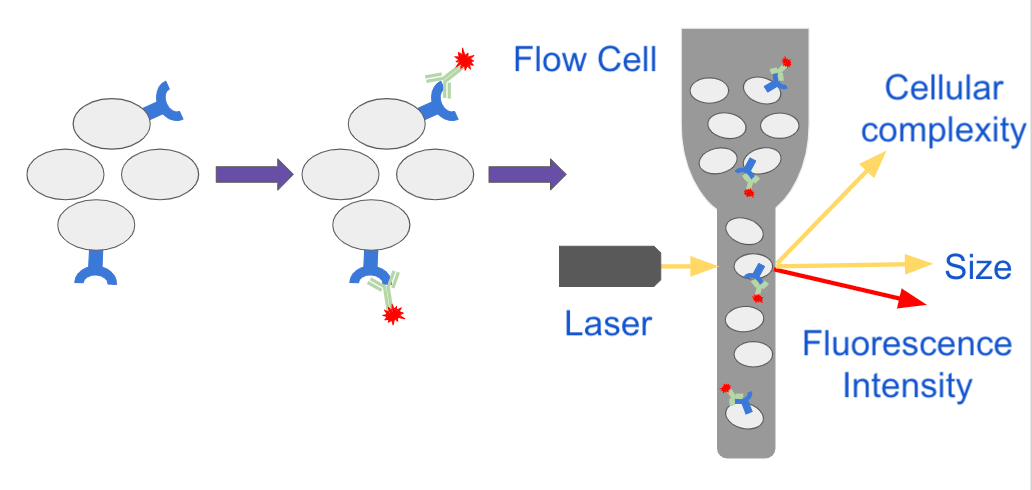
Analyzing Single Cells With Flow Cytometry
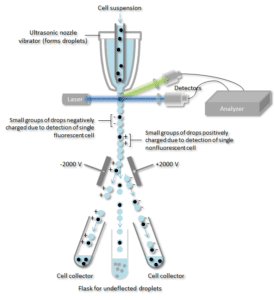
How Does Flow Cytometry Work Nanocellect

Optimized Flow Cytometric Protocol For The Detection Of Functional Subsets Of Low Frequency Antigen Specific Cd4 And Cd8 T Cells Sciencedirect

Fluorescence Activated Cell Sorting Of Live Cells Abcam

Fluorescence Activated Cell Sorting Facs Sino Biological
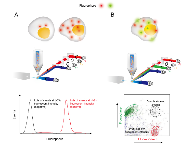
Flow Cytometry Guide Creative Diagnostics

Flow Cytometry Platform For Intracellular Detection Of Fviii In Blood Cells A New Tool To Assess Gene Therapy Efficiency For Hemophilia A Molecular Therapy Methods Clinical Development

The Principle Of Flow Cytometry And Facs 1 Flow Cytometry Youtube

Flow Cytometry Introduction Abcam

Workflow For Establishing The Fit For Purpose Of A Flow Cytometry Download Scientific Diagram

The Principle Of Flow Cytometry And Facs 1 Flow Cytometry Youtube
